


Science, Space and Technology News 2024
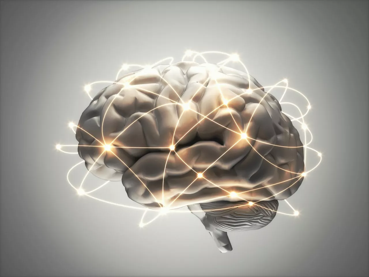
Scientists have collaborated to create the world’s first 3D-printed “brain phantom,” utilizing a special magnetic resonance imaging technique to model brain fibers. This advancement is aimed at improving research into neurodegenerative diseases such as Alzheimer’s, Parkinson’s, and multiple sclerosis by enhancing the accuracy of dMRI analysis software through the use of these detailed brain models.
In a joint project between MedUni Vienna and TU Wien, the world’s first 3D-printed “brain phantom” has been developed, which is modelled on the structure of brain fibres and can be imaged using a special variant of magnetic resonance imaging . As a scientific team led by MedUni Vienna and TU Wien has now shown in a study, these brain models can be used to advance research into neurodegenerative diseases such asMagnetic resonance imaging is a widely used diagnostic imaging technique that is primarily used to examine the brain. MRI can be used to examine the structure and function of the brain without the use of ionizing radiation. In a special variant of MRI, diffusion-weighted MRI , the direction of the nerve fibers in the brain can also be determined. However, it is very difficult to correctly determine the direction of nerve fibers at the crossing points of nerve fiber bundles, as nerve fibers with different directions overlap there. In order to further improve the process and test analysis and evaluation methods, an international team in collaboration with the Medical University of Vienna and TU Wien developed a so-called “brain phantom”, which was produced using a high-resolution 3D printing process.Researchers from the Medical University of Vienna as MRI experts and TU Wien as 3D printing experts worked closely with colleagues from the University of Zurich and the University Medical Centre Hamburg-Eppendorf. Back in 2017, a two-In the course of this, work was also carried out on brain phantoms as a use case together with the Medical University of Vienna and the University of Zurich. The resulting patent forms the basis for the brain phantom that has now been developed and is being supervised by TU Wien’s Research and Transfer Support team. Visually, this phantom does not have much to do with a real brain. It is much smaller and has the shape of a cube. Inside it are extremely fine, water-filled microchannels the size of individual cranial nerves. The diameters of these channels are five times thinner than a human hair. In order to imitate the fine network of nerve cells in the brain, the research team led by first authors Michael Woletz and Franziska Chalupa-Gantner used a rather unusual 3D printing method: two-photon polymerization. This high-resolution method is primarily used to print microstructures in the nanometre and micrometer range – not for printing three-dimensional structures in the cubic millimeter range. In order to create phantoms of a suitable size for dMRI, the researchers at TU Wien have been working on scaling up the 3D printing process and enabling the printing of larger objects with high-resolution details. Highly scaled 3D printing provides the researchers with very good models that – when viewed under dMRI – make it possible to assign various nerve structures. Michael Woletz compares this approach to improving the diagnostic capabilities of dMRI with the way a mobile phone camera works: “We see the greatest progress in photography with mobile phone cameras not necessarily in new, better lenses, but in the software that improves the captured images. The situation is similar with dMRI: using the newly developed brain phantom, we can adjust the analysis software much more precisely and thus improve the quality of the measured data and reconstruct the neural architecture of the brain more accurately.”The authentic reproduction of characteristic nerve structures in the brain is therefore important for “training” the dMRI analysis software. The use of 3D printing makes it possible to create diverse and complex designs that can be modified and customized. The brain phantoms thus depict areas in the brain that generate particularly complex signals and are therefore difficult to analyze, such as intersecting nerve pathways. In order to calibrate the analysis software, the brain phantom is therefore examined using dMRI, and the measured data is analyzed as in a real brain. Thanks to 3D printing, the design of the phantoms is precisely known and the results of the analysis can be checked. MedUni Vienna and TU Wien were able to show that this works as part of the joint research work. The phantoms developed can be used to improve dMRI, which can benefit the planning of operations and research into neurodegenerative diseases such as Alzheimer’s, Parkinson’s, and multiple sclerosis. Despite the proof of concept, the team still faces challenges. The biggest challenge at the moment is scaling up the method: “The high resolution of two-photon polymerization makes it possible to print details in the micro- and nanometre range and is therefore very suitable for imaging cranial nerves. At the same time, however, it takes a correspondingly long time to print a cube several cubic centimeters in size using this technique,” explains Chalupa-Gantner. “We are therefore not only aiming to develop even more complex designs, but also to further optimize the printing process itself.” Reference: “Toward Printing the Brain: A Microstructural Ground Truth Phantom for MRI” by Michael Woletz, Franziska Chalupa-Gantner, Benedikt Hager, Alexander Ricke, Siawoosh Mohammadi, Stefan Binder, Stefan Baudis, Aleksandr Ovsianikov, Christian Windischberger and Zoltan Nagy, 07 January 2024,SciTechDaily: Home of the best science and technology news since 1998. Keep up with the latest scitech news via email or social media.Toba supereruption may have facilitated the dispersal of modern humans out of Africa and across the rest of the world. Modern humans dispersed from Africa…
United States Latest News, United States Headlines
Similar News:You can also read news stories similar to this one that we have collected from other news sources.
 China: Scientists develop sensor to detect chemical weapons remotelyChinese scientists have developed a revolutionary new sensor to detect and monitor chemical weapon agents remotely.
Read more »
 Scientists Develop Potential New Method To Halt Progression of Alzheimer’s DiseaseScience, Space and Technology News 2024
Read more »
 Scientists Develop Optical Disc with Petabit Storage CapacityScientists have created a new type of optical disc, called AIE-DDPR, that can store up to 125 terabytes of data, equivalent to 15,000 DVDs. This new material offers a higher storage capacity than traditional HDDs and has the potential to revolutionize data storage.
Read more »
 Scientists Develop Technology to Make High Resolution Images of Human Spinal Cord During SurgeryScientists can make high resolution images of the human spinal cord during surgery with the help of functional ultrasound imaging (fUSI) technology. This advancement could provide relief to millions suffering from chronic back pain.
Read more »
 Scientists develop a rapid gene-editing screen to find effects of cancer mutationsResearchers found a way to screen cancer-linked gene mutations much more easily and quickly than existing approaches, using a variant of CRISPR genome-editing known as prime editing.
Read more »
 As scientists find real exoplanets, sci-fi writers change their vision of alien worldsKeith Cooper is a freelance science journalist and editor in the United Kingdom, and has a degree in physics and astrophysics from the University of Manchester.
Read more »